
Received: Sun 24, Aug 2025
Accepted: Wed 10, Sep 2025
Abstract
Objective: Laparoscopic inguinal hernia repair has been widely adopted as a primary treatment modality by numerous surgical centers due to its low rates of postoperative complications and recurrence. The present study aims to compare two distinct laparoscopic techniques- TEP and eTEP- in terms of technical feasibility, early postoperative quality of life, pain control, complication profile, and early recurrence rates.
Materials and Methods: Patients with an indication for laparoscopic inguinal hernia repair were randomly assigned to undergo either the TEP or eTEP procedure during July and August 2023. Postoperative follow-up included assessment of pain intensity at 24 hours using the Visual Analogue Scale (VAS) and evaluation of recovery quality using the quality of recovery test (QoR-15). At the first postoperative week, patients were reassessed for pain via VAS scoring, and physical examination was performed to identify any complications or early recurrence. Additional follow-up visits were scheduled for the first and third postoperative months.
Results: The TEP group demonstrated a significantly higher mean number of fixation staples (p < 0.001). Mean operative time was notably shorter with the eTEP approach (p = 0.031). Time to return to occupational activities was significantly prolonged in the TEP cohort (p = 0.024). On postoperative day one, VAS pain scores were markedly lower in the eTEP group (p < 0.001), whereas no statistically significant difference was observed between the groups at the first week. Similarly, QoR-15 scores at 24 hours post-surgery were significantly higher among patients undergoing eTEP (p = 0.006). No significant intergroup difference was detected regarding the incidence of seroma, ecchymosis, or hematoma.
Conclusion: While both TEP and eTEP techniques exhibited comparable complication rates, the eTEP method demonstrated potential advantages over TEP in terms of reduced operative time, enhanced early postoperative quality of life, and diminished acute postoperative pain.
Keywords
Extended total extraperitoneal repair, inguinal hernia, laparoscopic inguinal hernia repair, total extraperitoneal repair
Highlights
• The eTEP method offers an effective alternative to the commonly used TEP approach in laparoscopic inguinal hernia repair.
• eTEP reduces operation time due to its wider field of view and greater range of motion for instrument manipulation.
• eTEP is found to be more beneficial than TEP in terms of early acute postoperative pain and quality of life.
1. Introduction and Aim
Inguinal hernia is one of the most common disorders requiring referral to surgeons. Surgical repair is one of the most common methods in adults [1]. Today, many hernia repair methods are applied. With the recent use of synthetic patches and optical devices, this journey has reached laparoscopic hernia repair. Despite different surgical methods, the most important factors determining success are the absence of recurrence and chronic pain. There are extensive studies showing that failures have decreased considerably after the introduction of patches and laparoscopy [2, 3]. Laparoscopic approach is currently accepted as the first-line treatment in inguinal hernia repair [4]. These are generally TEP, transabdominal preperitoneal repair (TAPP), intraperitoneal onlay mesh repair (IPOM), primary repair and ring closure which can be performed in Nyhus type I hernias and in children. ETEP was developed by modification of TEP rather than a separate technique [5].
Favorable outcomes of laparoscopic surgery:
• Postoperative pain is less than open methods and the need for painkillers is less.
• Relatively low length of hospital stay.
• Early return to work.
• Fast wound healing.
• Providing more advantageous and reliable repair in recurrent hernias.
• Being more reliable and practical in bilateral hernias.
• Providing tension-free repair by including preperitoneal hernia repair and other risky possible hernia sites.
• In addition to hernia surgery in TAPP repair, other intra-abdominal pathologies can be checked in addition to hernia surgery.
• Cosmetic effects can be counted as.
Challenges of laparoscopic surgery:
• The need for more experience and anatomical knowledge compared to open surgery.
• Expensive costs.
• Need for laparoscopic courses and some clinical experience.
• TAPP repair has increased likelihood of morbidity.
• No significant difference in terms of recurrence has been demonstrated yet.
In this clinical study, TEP and eTEP methods were compared and operation times, postoperative quality of life and pain management, complication and early recurrence rates were evaluated.
2. Materials and Method
Our study, which was initiated with the approval of Fatih Sultan Mehmet Training and Research Hospital Clinical Research Ethics Committee on 13.07.2023 with the number KAEK 2023/102, was planned to be prospective and randomised. A total of 75 patients were included in the study to be operated between July and August 2023. Patients who were previously diagnosed with inguinal hernia in the general surgery outpatient clinic and planned surgery were included in the study according to the determined criteria.
Inclusion Criteria:
• Over 18 years of age.
• Patients who agreed a laparoscopic hernia repair.
• Unilateral inguinal hernia with fascia defect larger than 0.5 cm.
• EHS Classification (Primary, lateral or medial, 1, 2 and 3).
• ASA 1-3 (not in need of intensive care).
Exclusion Criteria:
• Bilateral hernias.
• Strangulated hernias.
• Patients in whom laparoscopic operation is contraindicated.
• Patients who developed recurrence after TEP or eTEP.
• Patients with a history of previous open lower abdominal operation.
• Patients who should not receive general anaesthesia.
• ASA 3 patients in need of intensive care and ASA 4 patients.
Patients were informed with a detailed voluntary consent form explaining the purpose and content of the study and their signed consent was obtained. Simple randomisation was applied for randomisation and computer-assisted randomisation (Link) website was used. For two different groups, two separate randomised number sequences were obtained and the order of which cases were to be operated with which technique was made according to this numbers. A random number sequence was obtained using a computer-aided system for randomisation and assignment of cases to which operation group. The cases were numbered and assigned to groups.
Age, gender, height, preoperative weight, body mass index (BMI), presence of diabetes mellitus and hypertension, smoking status, side of hernia, hernia defect and size of hernia bulge were recorded preoperatively. During the operation, the presence of direct, indirect or combined (direct and indirect together) hernia, number of mesh fixing staples (tacker) used, operation time (min), amount of bleeding (ml), peritoneal defect whether it occurred or not was recorded.
In all patients, BARD 3Dmax anatomical mesh was used and COVIDIEN Protack tacker was used as fixative. In the postoperative period, the patients' need for parenteral analgesia, postoperative hospital stay (days), postoperative day 1 VAS (Appendix 1) values and day 1 QoR 15t (Appendix 2) scores were recorded. PAROL drug with paracetamol active ingredient was administered as IV analgesic if the patient needed it.
Patients were discharged on the 1st postoperative day if there was no obstacle for discharge and called for control one week later. In the first week control, VAS score was questioned again and the presence of seroma, haematoma, CAE, early hernia recurrence was checked by physical examination and recorded. Afterwards, patients were called for control in the first and third months after surgery, and in case of pathology, it was noted and necessary examinations were requested.
2.1. Surgery Techniques; TEP Repair
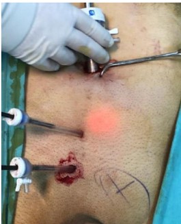
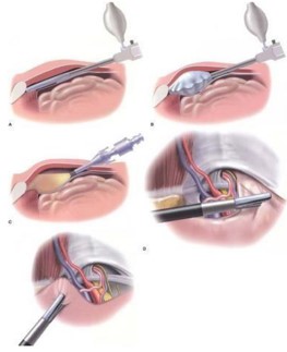
A 1.5 cm incision is made under the umbilicus and the anterior fascia of the rectus muscle is exposed. An incision is made in the anterior rectus sheath with a scalpel. The rectus muscle fibres are lateralised and the balloon trocar is advanced from the midline to the symphysis pubis through the posterior rectus sheath. Peritoneal space is bluntly dissected by inflating the balloon, and a 10 mm camera trocar is advanced to provide a low-pressure airspace. A low- pressure pneumoperitoneum of 9-11 mmHg is created to avoid subcutaneous emphysema. For appropriate imaging, the patient must have been given muscle relaxants by the anaesthesia team. The patient is given a 30-degree trendelenburg position and a 30-degree lateral position towards the side without hernia [6].
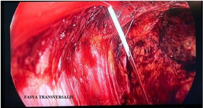
Then two 5 mm working trocars are inserted, one approximately 1 cm above the symphysis pubis and the other below the umbilicus, centring the camera trocar and the lower trocar as high as possible. The Cooper ligament on the hernia side is found and the preperitoneal fatty tissue attached to this ligament is bluntly dissected. The hernia area is seen during dissection. In these cases, the edge of the thinned fascia transversalis appears as a white border line because there is a pseudosac adherent to the preperitoneal fat. Lateral dissection is performed to create the Bogros cavity. The aim is to create the necessary space to place the prosthetic patch to be used. It is important to visualise the epigastric vessels and to keep them in the anterior abdominal wall. Since the vas deferens is a useful indicator in cord dissection, it is useful to find the spermatic vessels and vas deferens in cord dissection.
The peritoneum, called the hernia sac, is bluntly and sharply dissected from the spermatic cord and lowered as far into the abdomen as possible. After that, the mesh is shrunk in the dissection area and dropped into the cavity by means of the camera trocar. The mesh is placed to cover the hernia areas in accordance with its direction. Then the first fixation is stapled to the Cooper ligament. Afterwards, if necessary, the second tacker can be placed by paying attention to the epigastric vessels to re-fix the mesh to the anterior abdominal wall. It is not recommended to place a tacker lateral to the epigastric vessels to avoid damage to the lateral femoral cutaneous nerve, genitofemoralis nerve and ilioinguinal nerve.
After the mesh is fixed, the gas is desufflated while keeping the mesh fixed to the anterior abdominal wall. Since the TEP approach does not disrupt the peritoneal integrity, the risk of iatrogenic bladder and bowel damage is low. The fact that the peritoneum is not damaged reduces the possibility of contact of the mesh with intra-abdominal organs [6].
2.2. ETEP Repair
After marking the skin 3 cm above and 5 cm lateral to the umbilicus, a 1.5 cm incision is made and the anterior fascia of the rectus muscle is exposed. An incision is made in the anterior rectus sheath with a scalpel. The rectus muscle fibres are medialised and the posterior rectus sheath is seen and the balloon trocar is advanced to the lateral symphysis pubis. After by inflating the balloon, a 10 mm camera trocar is advanced through the incision. A preperitoneal air cavity with a low pressure of 9- 12 mmHg is created. In order to ensure a good visualisation, the patient should have full muscle relaxant drug efficacy. The patient is given same position towards the side without hernia [6].
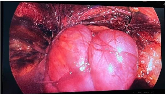
Two 5 mm working trocars are inserted, one from the midline just below the umbilicus and the other from slightly contralateral to the midline so that the symphysis pubis and umbilicus are in the middle. The Douglas band formed by the fascia transversalis, which is preferably seen just in front of and below the camera trocar, can be cut with scissors to provide better visualisation. In the presence of direct hernia, the hernia area is seen during dissection. Lateral dissection is also performed and Bogros cavity is created.
The peritoneum continuing on the cord as the hernia sac is dissected sharply and bluntly from the spermatic cord and reduced. Then, an appropriate mesh is shrunk and placed into the cavity through the camera trocar. The first fixation is stapled to the Cooper ligament. The second tacker can then be fixed medial to the epigastric vessels to fix the mesh to the anterior abdominal wall. After the mesh is fixed, the gas is desufflated.
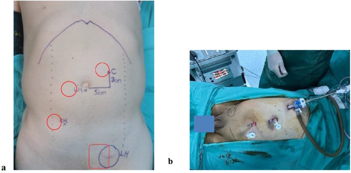
C: Port for Camera; LH: Port for Left Hand; RH: Port for Right Hand; LH: Left Inguinal Hernia.
2.3. Statistical Methods
Statistical analysis of all data obtained at the end of the study was performed with SPSS-27 (Chicago, USA) statistical programme. Normally distributed continuous variables were expressed as mean±standard deviation (SD) and non-normally distributed variables were expressed as median (interquartile range, 25-75%). Categorical variables were expressed as number and percentage. Chi-square test (Pearson ChiSquare) and Fischer Exact Test were used for the relationship between categorical variables. The parametric or nonparametric distributions of continuous variables were analysed by Kolmogorov-Smirnov test. Paired sample t-test was used to evaluate the differences between two groups in parametric variables and Mann Whitney-U test was used for nonparametric variables. For statistical significance, p ˂ 0.05 was accepted.
3. Results
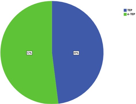
The study was conducted in the General Surgery Clinic of Fatih Sultan Mehmet Training and Research Hospital between July 2023 and August 2023. Our prospective and randomised study was conducted in 78 patients. And the data of 75 patients were analysed. Data collection and analysis were completed in November 2023. A total of 75 patients were studied, of which 36 underwent TEP and 39 underwent eTEP surgery (Figure 6).
The ages of the patients ranged between 33 and 85 years with a mean age of 57.5 years. Of the total 75 patients, 70 were male and 5 were female. BMI of the patients ranged between 18.90 kg/m² and 35.60 kg/m² with a mean value of 26.18 kg/m². When we analysed the comorbidities of the patients, we found diabetes in 8 (10.7%) patients and hypertension in 17 (22.7%) patients. 30 (40%) patients had smoking habit (Table 1). The mean ages of the TEP and e-TEP groups were 52.9±10.9 and 61.7±12.5 years, respectively, which were statistically significant (p:0.002) but not clinically significant (Table 3).
Table. 1: Demographic and other characteristics of all
patients (n=75).
|
Age |
|
|
Mean (±SD) |
57.5 (±12,4) |
|
Gender, n(%) |
|
|
Male |
70 (%93.3) |
|
Female |
5 (%6.7) |
|
BMI (kg/m²)(±SD) |
26.18 (±3.38) |
|
Diabetes Mellitus, n(%) |
8 (%10.7) |
|
Hypertension, n(%) |
17 (%22.7) |
|
Smoker, n(%) |
30 (%40) |
|
Side of operation, n(%) |
|
|
Right |
45 (%60) |
|
Left |
30 (%40) |
|
Direct/Indirect/direct+indirect, n(%) |
|
|
Direct |
9 (%12) |
|
Indirect |
45 (%60) |
|
Direct+ Indirect |
21 (%28) |
|
Defect (cm) |
|
|
Median (25-75%) |
1.50 (1.1-2.3) |
|
Diameter of swelling (cm²) |
|
|
Median (25-75%) |
4.0 (2.0-9.0) |
|
Number of Tacklers |
|
|
Median (25-75%) |
3.0 (2-4) |
|
Operation time (min) |
|
|
Median (25-75%) |
45 (35-55) |
|
Bleeding (ml) |
|
|
Median (25-75%) |
5 (0-5) |
|
IV Analgesic requirement, n(%) |
|
|
1 time |
14 (%18.7) |
|
2 times |
59 (%78.7) |
|
3 times |
2 (%2.7) |
|
Duration of hospitalisation (days) |
|
|
Median (25-75%) |
1.0 (1-1) |
|
Return to work time (days) |
|
|
Median (25-75%) |
8 (7-10) |
|
VAS day 1 score |
|
|
Median (25-75%) |
1.0 (0-2) |
|
VAS Week 1 score |
0 (0-1) |
|
Median (25-75%) |
0 (0-5) |
|
QoR15T |
|
|
Median (25-75%) |
128 (116-138) |
|
Seroma, n(%) |
4 (%5.3) |
|
Haematoma, n(%) |
1 (%1.3) |
|
Ecchymosis, n(%) |
7(%9.3) |
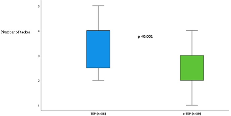
The hernia defect size of all patients ranged between 0.8 and 6.0 cm and the median defect size was 1.50 cm. The diameter of the bulge in the hernia area ranged between 1.0 and 36 cm² and the median value was 4.0 cm². When both groups were compared in terms of defect diameter and bulge diameter, there was no statistically significant difference between them. The number of tacker used varied between 1 and 5 and the median value was 3. When the number of tacker was compared, a highly significant difference was found between them (p<0.001) (Figure 7).
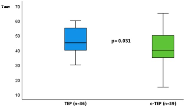
When both patient groups were compared in terms of gender, BMI, presence of diabetes mellitus, presence of hypertension, smoking and hernia sides, there was no statistically significant difference between the groups (Table 3). The operation time varied between 15 and 65 minutes in all patients and the median time was 45 minutes. We found a statistically significant difference was observed (p: 0.031) (Figure 8).
The amount of intraoperative bleeding ranged between 0 and 50 ml and the median amount of bleeding was calculated as 5 ml. However, no significant statistical difference was found. Assuming that a patient who needed one dose of paracetamol had mild pain and a patient who needed two or three paracetamol had moderate pain, 1 of 36 patients in the TEP group and 13 of 39 patients in the eTEP group had mild pain. In addition, 35 patients in the TEP group and 26 patients in the eTEP group had moderate pain (Table 2). Statistical comparison revealed a highly significant difference (p < 0.001) (Figure 9).
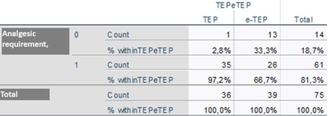
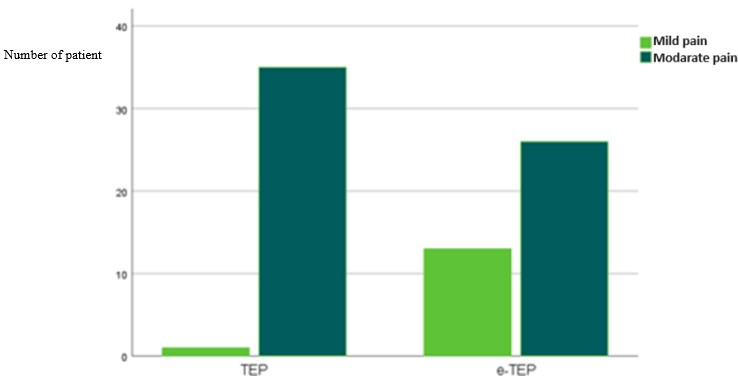
Table. 3. Comparison of demographic and other
characteristics in the TEP and e-TEP groups.
|
|
TEP (n=36) |
e-TEP (n=39) |
P |
|
Age |
52.9 (±10.5) |
61.7 (±12.5) |
0.002ᵃ |
|
Gender, n(%) |
|
|
0.18ᵇ |
|
Male |
32 (89) |
38 (97) |
|
|
Woman |
4 (11) |
1 (3) |
|
|
BMI, kg/m² (±SD) |
26.3 (2.6) |
26.05 (3.9) |
0.72ᵃ |
|
Diabetes Mellitus, n (%) |
4 (11) |
4 (10) |
1.0ᵇ |
|
Hypertension, n (%) |
5 (14) |
12 (31) |
0.08ᶜ |
|
Smoker, n (%) |
17(47) |
13 (33) |
0.22ᶜ |
|
Side of operation, n (%) |
|
|
|
|
Right side |
21 (58) |
24 (62) |
0.77ᶜ |
|
Left side |
15(42) |
15(38) |
|
|
Duration of surgery, median (25-75%) |
45(40-55) |
40 (35-50) |
0.031 ͩ |
|
Defect (cm), median (25-75%) |
1.65 (1.1-2.5) |
1.5 (1.1-2.1) |
0.82 ͩ |
|
Diameter of swelling (cm), median (25%-75) |
4.0 (2-9) |
4.0 (2-9) |
0.50 ͩ |
|
Number of tacklers, median (25-75%) |
4 (2.25-4) |
2 (2-3) |
<0.001 ͩ |
|
Bleeding (ml), median (25-75%) |
5 (0-10) |
0 (0-5) |
0.44 ͩ |
|
Seroma, n (%) |
1 (2.9) |
3 (7.7) |
0.61ᵇ |
|
Ecchymosis, n (%) |
4 (11) |
3 (8) |
0.70ᵇ |
|
Duration of hospitalisation (days), median (%25-75) |
1 (1-1) |
1(1-1) |
0.27 ͩ |
|
Return to work time (days), median (%25-75) |
8 (8-10) |
7 (7-9.5) |
0.024 ͩ |
|
VAS Day 1 score, median (25%-75) |
2 (0-2) |
0 (0-1) |
<0.001 ͩ |
|
VAS 1st week score, median (25%-75) |
1 (0-1) |
0 (0-1) |
0.057 ͩ |
|
QoR15T, median (25-75%) |
125 (107-1319 |
131(123-141) |
0.006 ͩ |
QoR15T: Quality of Recovery 15 Question Test;
SD: Standard Deviation; VAS: Visual Pain Scoring; BMI: Body Mass Index.
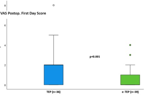
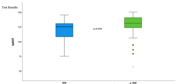
The postoperative hospitalisation period was generally 1 or 2 days and the median value was calculated as 1 day. It was calculated that there was no significant difference. The return to work time varied between 7 and 12 days and the median return to work time was 8 days. We found significant statistical difference (p: 0.024). In the postoperative VAS scores of the patients; 1st day scores were between 0 and 8 with a median score of 1, and 1st week scores were between 0 and 5 with a median score of 0. When both groups were compared, although there was a highly significant difference between the 1st day scores (p<0.001), there was no statistically significant difference in the 1st week scores (Figure 10). When we looked at the QoR 15T results, total scores ranged between 57 and 150 and the median value was calculated as 128. A significant statistical difference was found in the comparison of both groups (p=0.006) (Figure 11).
Seroma developed in 4 patients (5.3%) in the 1st week postoperatively and was aspirated after ultrasonography. The seroma regressed in the follow-up. Only 1 patient (1.3%) developed haematoma and no intervention was required. At the 3rd month follow-up, complete regression was observed. Ecchymosis was observed in the groin and scrotal region in 7 patients (9.3%) at the 1st week postoperative control, but no evidence of ecchymosis was found at the 1st month control of these patients. No significant difference was found.
4. Discussion
Inguinal hernia repair is one of the most common elective operations in general surgical practice. The development of minimally invasive surgical methods has raised the question of which method is the best in inguinal hernia operations. In TAPP, the surgeon enters the peritoneal cavity and places a mesh over possible hernia sites through a peritoneal incision. The difference of TEP is that the peritoneal cavity is not entered and mesh is used to close the hernia from outside the peritoneum. Since the peritoneal cavity is not entered, it is advantageous in terms of reducing the risk of intestinal or vascular injury and preventing trocar site hernia. Moreover, in some patients the procedure can be performed under regional anaesthesia with intravenous sedation.
Surgeons interested in laparoscopic surgery should be successful in all techniques so that they can tailor their approach to the needs of the patient. TEP has shown several flaws that surgeons have struggled to overcome over the years. These include limited space for dissection and the associated difficulty in spreading the patch, cramped port placement, possible intolerance to pneumoperitoneum, and most importantly, TEP has a long learning curve for young surgeons. Considering the above-mentioned conditions, the eTEP method was designed by Jorge Daes by modifying the TEP method. The most prominent features of this method are rapid and easy creation of the extraperitoneal space, a large surgical field, a flexible port setup, free parietalisation of the spermatic cord (dissection of the proximal hernia sac and peritoneum), easier management of the distal hernia sac in large inguino-scrotal hernias, and increased tolerance to pneumoperitoneum.
Daes determined that the eTEP method is more usable in certain patient groups and situations. These are; obese and postbariatric patients, who may be preferred by novice surgeons with no experience in TEP repair, patients with short distance between pubic tubercle and umbilicus, patients with a history of previous pelvic surgery, and patients with giant inguinal hernia [6]. When we look at the literature published to date, the number of randomised clinical studies related to eTEP is small. It was generally prospectively compared with other methods. In this work, we tried to demonstrate which method is more comfortable for the patient and easy to be applied by surgeons with different parameters by applying different methods.
In our study, the mean ages of the TEP and eTEP groups were 52.9±10.9 and 61.7±12.5 (p: 0.002), respectively. Although a statistically significant difference was found between the mean ages of the two groups, it was not considered as a clinically significant difference. In daily practice, this age difference was interpreted as a negligible age difference in routine surgical operations. In addition, there are not enough studies in the literature showing that there may be a serious difference between the 50 and 60 age group in terms of surgical complications (hospitalisation time, bleeding, revision leakage, etc.). As expected, since the sample group was small, it is highly likely that such differences may occur due to the order during randomisation. In the study published by Köckerling et al. in which TEP and TAPP methods were compared, the mean ages of the groups were 58.3 and 59.2 years, which overlapped with the mean age of the patients in our study [7].
And also, the mean BMI of the patients was 26.18 and similar values were observed in similar studies. As seen in the same studies, we found that there was no significant difference between our groups in terms of comorbidities and differently, we also questioned smoking which affects wound healing as much as obesity and comorbidities [8]. In some cases, a larger number of fixation staples used in patients are needed. For example, defect diameter and bulge diameter are among the parameters affecting this situation. Although there was no significant difference between the groups in terms of these two parameters in our study, the number of staples needed in the eTEP group were significantly less. In the study published by Reza et al., patients in whom eTEP method was applied were analysed and it was shown that 2 staples were needed in 60% of the patients and single staple was used in 28% [8].
In the study published by Singh et al., TEP and eTEP methods were compared in patients and it was shown that 1 or 2 staples were used in the TEP method, whereas the number of staples needed varied between 4 and 6 in patients who underwent eTEP method [9]. In many studies, it has been shown that the number of staples used is correlated with postoperative pain. In our study, the fact that fewer staples were needed in the eTEP group suggests that patients may have a more comfortable postoperative period. There is a strong correlation between the ease of dissection and the duration of surgery. The study revealed that the mean operation time was significantly shorter in the eTEP group. In a similar study, it was shown that the eTEP group had a significantly shorter mean operation time [8]. However, although the mean operation times of the groups in this study were 167.6 ± 32.4 and 127.5 ± 23.4 min, we showed that the operation time ranged between 15 and 65 min and the median time was 45 min in our patients.
Peritoneal rupture occurred in a total of 2 patients during the operation, one each in the TEP and eTEP groups. None of them required verses decompression or peritoneal defect closure with loop. There was no need to switch to another method in any patient. In the current studies, patients were evaluated in terms of parameters such as peritoneal rupture and exposure, but no significant difference was observed. However, in a study which TEP and eTEP were compared, transition to TAPP method was made in 4 patients in the TEP group because the peritoneal defect could not be controlled (p:0.037) [8].
In laparoscopic extraperitoneal inguinal hernia operations, bleeding usually occurs from capillaries in the lateral wall, spermatic cord vessels, vascular structures around the ramus pubis and sometimes from the inferior epigastric artery and vein. In most of the studies, the TEP method was compared to other laparoscopic methods. Although it provided a better working area and less intestinal damage, the amount of bleeding was significantly higher than that of the previous study [10]. The reason for this has been shown to be that TEP provides a more limited area in the management of bleeding [11]. In our study, we found no significant difference between the groups in terms of the amount and incidence of bleeding. We easily controlled significant bleeding with vessel sealing devices. We did not place drains in the patients, we followed our patients, only if necessary, with a serum bag placed on the pubic region postoperatively and removed it when the patients started to walk on the evening of the operation. We did not find subcutaneous emphysema in the operation area in any of the patients.
We did not use NSAI analgesics in the management of pain, and we only used IV paracetamol in a number ranging from 1-3. Although we found that 2 doses with an 8-hour interval were sufficient in most of the patients (78.7%), we found that the need for more than one dose was statistically significantly higher in the TEP group compared to the other group. At the studies published so far; in a study comparing TEP and TAPP, the groups were evaluated with VAS and no significant difference was found between them in terms of pain [7]. In another study, patients who underwent eTEP were evaluated with VAS at the 1st week. While 60% of the patients had no pain, 32% had VAS scores of less than 4. In a study comparing TEP and eTEP methods, pain scores were evaluated with VAS from the day of operation to the 1st week control and it was found that pain scores were significantly lower in the eTEP group [8].
In our study, 1st day VAS scores were between 0 and 8 in all patients (median value 1) and pain scores were significantly lower in the eTEP group. This result may be associated with the fact that the need for only 1 dose of paracetamol was significantly higher in the eTEP group compared to the other group. However, in contrast to the other study, although our sample size in the groups was larger, there was no significant difference in 1st week pain scores. None of the patients had shoulder pain, which has been observed in some patients in other similar studies. In the TEP group, more field dissection was required with laparoscopic instruments, whereas in the eTEP group, the balloon required more dissection. With eTEP we dissected more area effectively. This may explain the lower pain scores on postoperative day 1 in the eTEP group. All patients were prescribed prophylactic antibiotics and NSAI tablets at discharge, and the patients used the painkiller tablets as needed. Therefore, we found that the median value of the VAS scores at the 1st week control was 0 and there was no significant difference between the groups.
General surgeons mostly use the Short Form 36 (SF36) test to evaluate the quality of life of postoperative patients. In a study conducted by Lawrence et al., patients who underwent inguinal hernia repair by open and laparoscopic methods were evaluated with SF-36 questionnaire at the 3rd and 6th months, but no significant difference was found between the groups [11]. QoR 15 test is a questionnaire questioning the general condition of the patients except for pain, which has started to be used frequently in studies performed with anaesthetised surgical patients. Overall, while the SF-36 serves as a board measure of health-related quality of life, the QoR-15 provides a more practical and sensitive tool for evaluating postoperative recovery. At the studies, we realised that it had not been used in patients who underwent inguinal hernia surgery, so we decided to include it in our study to evaluate the patients. We used the QoR 15 test revised in Turkish by Kara et al. We used the QoR 15t questionnaire revised in Turkish [12]. Only postoperative at 24 hours, these tests were collected and analysed for the two groups. When we looked at the results, we saw that significantly higher scores were obtained in the eTEP group.
The fact that the duration of the operation, the number of staples used, and the 1st day pain scores were significantly different between the two groups showed that there was a significant difference in postoperative quality of life. We recorded the postoperative hospitalisation time of each patient and the return to work time of working patients in days. We found that the return to work time was significantly less in the eTEP group, but there was no significant difference in the hospitalisation times. When we look at the current literature, Singh et al. compared the TEP and eTEP groups on 25 patients and found that the mean hospital stay was 1.7±0.7 days and 1.1±0.3 days (p < 0.001) in the TEP group, respectively. In this study, the mean return to work time between the groups was calculated as 11.6±3.6 days and 9.9±3.4 days, respectively (p: 0.093) and no significant difference was found between TEP and TAPP [9].
In the studies, it was found that the hospital stay was longer in the TAPP group and the return to work time was shorter in the TEP group in one study, but these differences were not significant [13]. In a study conducted in Switzerland with 1309 patients (287 of which were TAPP and the others were TEP), it was shown that the duration of hospital stay was significantly longer in the TAPP group, but the return to work time was not evaluated [14]. In a study comparing TAPP and eTEP methods, it was found that the length of hospital stay was significantly less in the eTEP group [15]. Today, it is accepted by many surgical institutions that prolonged hospital stay increases hospital-acquired complications and increases the financial burden in terms of both patient and hospital management. From this point of view, both methods are safe options. A short return to work time is also important for working patients to make their surgical plans more easily.
In our study, complications such as seroma, ecchymosis and haematoma development at the wound site were evaluated both during the hospitalisation process and at the 1st week, 1st month and 3rd month clinic controls. In the TEP group, 1 patient developed seroma, 4 patients developed inguinal and scrotal ecchymosis, and 1 patient developed haematoma in the inguinal region. In the other group, we observed seroma in 3 patients, ecchymosis in 3 patients and no haematoma. No statistically significant difference was observed in accordance with similar studies in the literature.
The most important indicator of successful hernia repair surgery is the non-recurrence of hernia. Today, the recurrence rate of inguinal hernia is still accepted as 10% by many surgical institutions. In our study, no early recurrence of inguinal hernia was observed in either group at the end of the 3-month follow-up; however this period is not sufficient to evaluate mid- and long-term recurrences. When we look at the current literature, most of them have follow-up periods ranging between 1 and 6 months and have not reported recurrence. In one study, patients who had previously undergone open hernia repair surgery but had recurrence were operated with TEP and TAPP methods. Patients were followed up for 1 year and recurrence was observed in 7 (0.90%) patients in the TEP group and 21 (1.43%) patients in the TAPP group, but no significant difference was found (p:0.275) [7]. In another study, TEP and eTEP methods were applied to patients in 2 years and compared and no recurrence was observed in any of the groups, but the mean follow-up period of the patients was not specified in the study [9].
5. Conclusion
Although the TEP method is one of the most commonly preferred methods in laparoscopic inguinal hernia repair, we think that the eTEP method, which has been developed later, can be easily preferred in patients. eTEP approach is advantageous compared to the TEP approach in terms of operation time due to its wider angle of view and wider range of motion for instrument manipulation. It was also found that eTEP was more advantageous than TEP method in early acute postoperative pain and quality of life. However, this study was conducted as a single-center clinical study, and larger multicenter studies are needed to confirm the findings.
Conflicts of Interest
None.
Funding
None.
Author Contributions
B. Agca (Corresponding author): Responsible for the overall study design and patient management; most of the patients included in the study were referred to him for surgery. Y. Gunes: Assisted in the postoperative follow-up of patients on the ward. O. Segmen: Contributed to the discussion section by helping with the literature review and referencing. M.T. Aydin: Allowed the inclusion and surgical treatment of a small number of patients who were referred to him. E. Onur: Similarly permitted the inclusion and surgical treatment of some of his referred patients. K. Memisoglu: Was the clinical education supervisor at the time and provided guidance on the study's methodology. All authors reviewed and approved the final manuscript.
Ethical Approval
The study was approved by the Ethics Committee of Fatih Sultan Mehmet Training and Research Hospital (No: KAEK 2023/102, Date: 13.07.2023).
Informed Consent
Informed consent was obtained from all participants included in the study.
Data Sharing and Accessibility
All data related to the patients are stored in physical files. The corresponding Excel files containing these data are available in our institutional library. Upon request, the data can be shared digitally.
Data Statement
The data generated and analyzed during this study are stored in Excel format at the institutional library of the Fatih Sultan Mehmet Training and Research Hospital. The data are not publicly shared but can be made available upon reasonable request from the corresponding author. Ethical approval for data sharing was obtained from the Ethics Committee of Fatih Sultan Mehmet Training and Research Hospital (No: KAEK 2023/102, Date: 13.07.2023). The data are strictly for research purposes and have not been used for commercial purposes.
REFERENCES
[1] W Y Lau “History of
treatment of groin hernia.” World J Surg, vol. 26, no. 6, pp. 748-759,
2002. View at: Publisher
Site | PubMed
[2] D Arvidsson, F H Berndsen,
L G Larsson, et al. “Randomized clinical trial comparing 5-year recurrence rate
after laparoscopic versus Shouldice repair of primary inguinal hernia.” Br J
Surg, vol. 92, no. 9, pp. 1085-1091, 2005. View at: Publisher Site | PubMed
[3] A Eklund, A Montgomery, L
Bergkvist, et al. “Chronic pain 5 years after randomized comparison of
laparoscopic and Lichtenstein inguinal hernia repair.” Br J Surg, vol.
97, no. 4, pp. 600-608, 2010. View at: Publisher Site | PubMed
[4] K McCormack, N W Scott, P
M Go, et al. “Laparoscopic techniques versus open techniques for inguinal
hernia repair.” Cochrane Database Syst Rev, vol. 2003, no. 1, pp.
CD001785, 2003. View at: Publisher
Site | PubMed
[5] B J Leibl, C Jäger, B
Kraft, et al. “Laparoscopic hernia repair--TAPP or/and TEP?” Langenbecks
Arch Surg, vol. 390, no. 2, pp. 77-82, 2005. View at: Publisher Site | PubMed
[6] Jorge Daes “The enhanced
view-totally extraperitoneal technique for repair of inguinal hernia.” Surg
Endosc, vol. 26, no. 4, pp. 1187-1189, 2012. View at: Publisher Site | PubMed
[7] F Köckerling, R Bittner, A
Kuthe, et al. “TEP or TAPP for recurrent inguinal hernia repair-register-based
comparison of the outcome.” Surg Endosc, vol. 31, no. 10, pp. 3872-3882, 2017. View at: Publisher Site | PubMed
[8] Syed Masud Reza, Md
Mozammel Hoque, SM Quamrul Akther, et al. “Early Outcomes of "Extended
View Total Extraperitonial (eTEP)" Procedure for Inguinal Hernia.” J
Shaheed Suhrawardy Med Coll, vol. 11, no. 2, pp. 96-100, 2019. View at: Publisher Site
[9] Siddharth Singh, Sanjay
Kala, Jauhari RK, et al. “A prospective randomized study of eTEP and TEP repair
for inguinal hernia in terms of ease of operability, complication and
recurrences.” Asian Journal of Medical Sciences, vol. 13, no. 3, pp.
87-94, 2022. View at: Publisher
Site
[10] Deborshi Sharma, Kamal
Yadav, Priya Hazrah, et al. “Prospective Randomised trial comparing
laparoscopic transabdominal preperitoneal (TAPP) and laparoscopictotally
extraperitoneal (TEP) approachfor bilateral inguinal hernias.” Int J Surg,
vol. 22, pp. 110-117, 2015. View at: Publisher Site | PubMed
[11] K Lawrence, D McWhinnie, C
Jenkinson, et al. “Quality of life in patients undergoing inguinal hernia
repair.” Ann R Coll Surg Engl, vol. 79, no. 1, pp. 40-45, 1997. View at:
PubMed
[12] Umut Kara, Fatih Şimşek,
Hasan Kamburoğlu, et al. “Linguistic validation of a widely
used recovery score: quality of recovery-15 (QoR-15). Turk J Med Sci,
vol. 52, no. 2, pp. 427-435, 2022. View at: Publisher Site | PubMed
[13] G Vinay, KS
Balasubrahmanya “Comparative study on Laparoscopic Transabdominal Pre-
Peritoneal(TAPP) Mesh Repair and Total Extra-Peritoneal(TEP) Repair in Inguinal
Hernia.” Madridge J Surg, vol. 1, no. 1, pp. 9-13, 2018. View at: Publisher Site
[14] M Gass, A Scheiwiller, M
Sykora, et al. “TAPP or TEP for Recurrent Inguinal Hernia? Population-Based
Analysis of Prospective Data on 1309 Patients Undergoing Endoscopic Repair for
Recurrent Inguinal Hernia.” World J Surg, vol. 40, no. 10, pp.
2348-2352, 2016. View at: Publisher
Site | PubMed
[15]
Kancham Nethaji, Rinku Kumari,
Pradeep Jaiswal, et al. “Comparison of Extended Total Extraperitoneal (E-TEP)
Repair and Trans-Abdominal Pre-Peritoneal (TAPP) Mesh Repair in Inguinal Hernia
Repair. Cureus, vol. 15, no. 5, pp. e39420, 2023. View at: Publisher
Site | PubMed