
Received: Fri 12, Dec 2025
Accepted: Wed 07, Jan 2026
Abstract
Introduction: Vanishing lung syndrome (VLS) is a relatively rare respiratory disease in clinical practice. The disease has a slow onset and is highly insidious, causing significant damage to the patient's respiratory function. In recent years, this disease has gradually received clinical attention. However, current clinical reports on it are mostly limited to clinical findings, diagnosis and treatment, and there are few reports on the long-term changes in lung damage caused by this disease. By understanding the long-term pulmonary imaging changes (i.e., pulmonary morphological changes) of VLS, clinicians can better grasp the evolution process of this disease, providing a basis for subsequent etiological analysis, diagnosis, and treatment.
Case Report: This paper reports the pulmonary imaging changes of a VLS patient for 12 years and the experience and lessons of treatment. A 40-year-old male patient had a history of type IV VLS for 10 years. He was diagnosed with "left spontaneous tension pneumothorax" this time and a partial lung bulla resection was performed in our department. After the surgery, the patient's clinical symptoms had significantly improved. However, on the third day, a continuous leakage of the pleural cavity occurred, causing widespread subcutaneous emphysema throughout the body. Adequate thoracic drainage was provided, some gas was retained in the pleural cavity to reduce the negative pressure in the lungs until the complication healed. The patient was cured and discharged. Two years later, a re-examination was conducted. The results found that surgery could increase the patient's effective breathing area relative to the previous operation. The patient's condition improved significantly. The chest CT scans of the patient over the past 12 years showed that in the early stage of the disease, the overall progression of lung damage was relatively rapid, but it gradually slowed down in the later stage.
Conclusion: This article presents the imaging changes of the lungs over a relatively long period of time, providing first-hand clinical data for analyzing the deterioration process of the disease and further understanding the pathological and physiological process of the disease. It also clarifies that VLS is the final clinical manifestation of a series of diseases, rather than the final diagnosis. It also proposes a new clinical classification scheme based on the cause.
Keywords
Vanishing lung syndrome, bullae of lung, imaging changes, surgical treatment
1. Introduction
The introduction should be succinct, with no subheadings. Limited figures may be included only if they are truly introductory, and contain no new results. Vanishing lung syndrome (VLS), also known as idiopathic giant bullous emphysema (GBE) [1], Type I bullous disease or idiopathic bullous disease [2], idiopathic large bulla [3], etc., is a relatively rare lung disease in clinical practice. Its pathological feature is large bullae of pulmonary emphysema, usually developing in the upper lung lobe and occupying at least one-third of a lung lobe. This is a long-term, progressive and irreversible disease involving destruction of lung parenchyma and expansion of alveoli. The effective respiratory area of the lungs gradually decreases [4]. It is usually associated with smokers, long-term marijuana users and methamphetamine users, HIV patients, and patients with α-1-antitrypsin deficiency. There have also been reports that it is a rare manifestation of chronic obstructive pulmonary disease (COPD) [5]. In addition, new research has found that this disease also has a tendency for familial inheritance [6]. It may also be closely related to the infection of the novel coronavirus (COVID-19) [7]. Rare causes include pulmonary nodule disease inducing large bullae to cause VLS, etc. [8].
VLS poses significant harm to the human body, often leading to fatal spontaneous pneumothorax [3], progressively worsening respiratory difficulties, hypoxemia [9], lung infections [10], and even uncontrollable bronchopleural fistula. Sometimes, even with surgical treatment, it is difficult to achieve a full recovery, and patients may be forced to have their affected lungs removed [11]. For some patients with VLS, the giant bullae in their lungs are close to the heart. Due to the compression of the heart by the huge bullae, patients may suddenly faint [12]. All these pose great risks to the patients' daily lives. Therefore, it is extremely important to deepen the understanding of the causes of VLS attacks, the pathophysiological process, as well as the pathological changes, clinical manifestations and diagnosis of the patients' lungs throughout the long-term disease course, and based on this, determine the treatment plan. Chest imaging examination, especially chest CT examination, plays the most important role [13]. Due to the aforementioned reasons, this paper reports on a VLS patient, presenting the changes in the lungs' CT scans over a period of 12 years, and evaluating the effectiveness of the surgical treatment.
2. Case Report
2.1. Clinical Manifestations and Medical History
Patient, male, 38-years old. Presented to the thoracic surgery department with symptoms of "shortness of breath accompanied by cough and yellow sputum for one week, and the condition worsened for two days". The aforementioned symptoms began a week ago. The patient treated them with antibiotics on their own, but the treatment was ineffective. Two days ago, the condition worsened and the patient experienced a significant increase in breathing difficulties. There is no history of smoking in the past. During the youth period, there was a history of tuberculosis in both lungs, which was cured after professional treatment. Ten years ago (at the age of 28), large bullae were found in both lungs and gradually worsened. There is mild breathing difficulty usually, and salbutamol inhalation aerosol is used for symptomatic treatment. During this period, there were multiple occurrences of pulmonary infections, which were all treated with corresponding antibiotics until recovery. No special drug treatment was given. Chest CT follow-up examinations were conducted approximately every two years. Currently, it is the tenth year since the discovery of VLS (at the age of 38). The patient stated that no one in the family had a history of similar lung diseases.
2.2. Physical Examination
On physical examination, the basic vital signs are as follows: body temperature 38.5°C, blood pressure 145/76 mmHg, heart rate 95/min, respiratory rate 30/min. Percutaneous blood oxygen saturation: 83%. The patient is conscious, breathing is rapid, and the lips are slightly cyanotic. The breath sounds in the right thoracic cavity are weakened, the breath sounds in the left thoracic cavity have disappeared, and expiratory crackles can be heard in the right lung, and a small amount of moist rales can be heard at the bottom of the right lung. Heart sounds can be heard, the rhythm is regular, no abnormal sounds in each valve are heard, but the strongest points of each valve's heart sounds are all bias on the right side. No abnormalities were found in the physical examination of the head and abdomen. The limbs are normal, and no edema is observed. The rest of the physical examination was normal.
2.3. Laboratory Examinations and Imaging Examinations
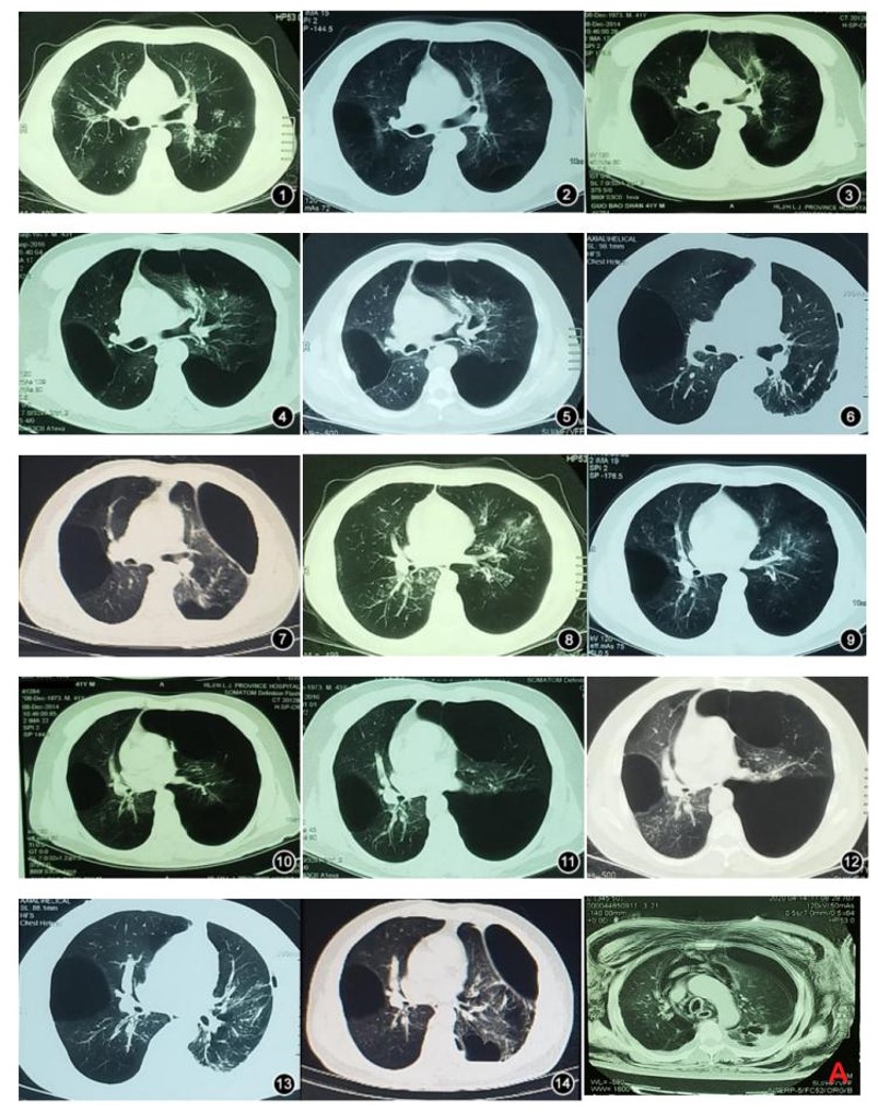
The blood routine test showed white blood cell count was 11.2×10⁹/L, with no other abnormalities such as anemia. The biochemical tests: liver function, kidney function, myocardial enzymes, various ions, blood lipids, etc. were all normal. Chest CT shows the patient's lung tissue content in both lungs is significantly reduced compared to normal lung tissue. Multiple bullae-like images can be seen in both lungs. A large amount of gas is visible in the left thoracic cavity, compressing the lungs and presenting as a tension pneumothorax. Patchy images can be seen in the remaining right lung. The volume of the right thoracic cavity is reduced, and the mediastinum is shifted to the right (Figure 1, ⑤ and ⑫
2.4. Diagnosis
According to the radiological diagnostic criteria proposed by Roberts et al. in 1987 [14], combined with the medical history and clinical examination results, the diagnosis was: left-sided tension pneumothorax, type IV VLS, pulmonary infection, and bronchospasm.
2.5. Treatment and Results
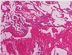
The patient was admitted to our department due to aggravated dyspnea and the failure of drugs to relieve hypoxia. A partial bulla resection of the left lung was performed successfully. A 9F closed thoracic drainage tube was placed during the operation. The pathology confirmed it to be bullous lung tissue (Figure 2). Antibiotics were used to assist in the treatment of pulmonary infection. The patient's initial postoperative recovery was good, with the following vital signs: body temperature 37.5°C, blood pressure 128/70 mmHg, heart rate 85 /min, respiratory rate 20/min. The transcutaneous oxygen saturation was 95%. The patient's clinical symptoms improved significantly. However, three days after the operation, the patient suddenly developed extensive subcutaneous emphysema in the chest wall, head and neck, and abdomen, as well as mediastinal emphysema (Figure 1A). The 9F drainage tube was continuously leaking gas. A re-examination of the chest CT showed tension pneumothorax on the surgical side.
Therefore, the 9F drainage tube was replaced with a 28F closed thoracic drainage tube for drainage, and the speed of gas drainage was controlled to maintain a certain amount of gas in the thoracic cavity to reduce the negative pressure in the thoracic cavity. After the subcutaneous emphysema disappeared, the drainage tube was attempted to be clamped, and some gas was retained in the thoracic cavity to reduce the negative pressure in the thoracic cavity. After repeatedly confirming that the patient had no adverse reactions, the drainage tube was removed. The patient was discharged after 42 days of recovery. In 2022, a follow-up re-examination was conducted. The chest CT showed that although the respiratory area of the lung on the surgical side of the patient was reduced compared to immediately after the operation, it was still significantly larger than before the operation. There was no significant change on the non-surgical side. The evolution of the patient's lung disease over the past ten years as seen on CT scans is shown in (Figure 1). Based on the CT results, the upper lobe of the left lung and the posterior segment of the lower lobe of the right lung, where the lung changes were more significant, were selected to observe the overall evolution of the lung disease.
At the same time, the results obtained in combination with the diagnosis and treatment are as follows: First, from the time trend, the overall progression of lung damage in the early stage of the patient's disease was relatively fast, but gradually slowed down later. The patient's condition progressed rapidly in the first six years and deteriorated significantly; in the next six years, the disease progressed slowly, especially in the right lung, where there was almost no significant progression, and the damage rate of the left lung was also relatively slow, but the overall trend was still deteriorating. Second, the lung damage in the patient was not uniform and showed asymmetry in time and location of onset. In this case, the deterioration rate and degree of the left lung were more obvious than those of the right lung. Third, within a certain range, surgical volume reduction treatment can increase the respiratory area of the lung and improve the patient's respiratory function. In the two years after the operation, the patient's condition was significantly improved compared to before the operation. However, when observed over a two-year period, the CT showed that the patient's condition was still slowly deteriorating, and the long-term efficacy still needs further observation. Fourth, from the clinical manifestations, the patient's lung resistance was poor and they were prone to repeated pulmonary infections.
2.6. Outcome and Follow-up
The patient's breathing difficulty has significantly improved, but still uses salbutamol inhalation aerosol for symptomatic treatment. The lungs still experience frequent infections that require treatment.
2.7. Figures
3. Discussion
3.1. The Current Status of VLS Diagnosis and Treatment
Although the incidence of VLS is relatively low, there are still numerous studies reporting on it. As far as the current literature is concerned, it mainly focuses on clinical findings, diagnosis, and treatment. A small amount of research has been conducted on the causes of the disease [6], genetics [7], and other factors. Moreover, almost all the schemes are surgical removal of pulmonary bullae and symptomatic drug treatment. This can only temporarily alleviate the condition and is not a true treatment for VLS. VLS is a long-term disease with gradually worsening symptoms [4]. The above treatments cannot fundamentally change the long-term pathological damage to the lung parenchyma and alveolar dilation and other pathological impairments. Therefore, even if patients undergo surgical treatment and/or drug treatment to temporarily alleviate some symptoms, they will inevitably eventually experience a gradual reduction in lung volume, a decrease in effective respiratory area, and ultimately respiratory failure. Therefore, finding the cause of VLS patients, observing the long-term pathological and pathophysiological changes of the lungs, and further deepening the understanding of this disease based on these changes, in order to find better treatment plans for the future, is the ultimate solution for treating VLS. At the same time, clinicians can diagnose VLS patients early by fully understanding the CT manifestations of their lungs at different time points and formulate corresponding treatment plans, which is of great significance.
3.2. Patient Characteristics
This paper tracked the natural evolution of the condition of this VLS patient for ten years and followed up for another two years after surgery, systematically monitoring the changes in the lungs of VLS patients on chest CT and the impact of minimally invasive surgery on the patients. Through the clinical manifestations of CT, we found that the destruction of the lung parenchyma and alveolar dilation in this VLS patient did not uniformly deteriorate over time; it presented a trend of being faster at first and then slower. The lesion sites were not as described in the previous literature, but rather multiple lung lobes simultaneously appeared with varying degrees of lesions at the same time [14, 15], which is different from the aforementioned literature [4]. The two-year follow-up after surgery showed that the surgery effectively improved the patient's clinical symptoms such as breathing difficulty and hypoxia, and these clinical manifestations were relatively stable. Studies had reported that VLS patients can maintain stable conditions within five years after surgery, and their lung function has improved [16]. This is consistent with the performance of this patient. Therefore, minimally invasive surgery to remove all or part of the pulmonary bullae is beneficial for patients [17-19].
However, for VLS patients, the surgery of pulmonary bulla resection carries certain risks. Due to the large size of the pulmonary bullae and the limited amount of functional lung tissue, postoperative drainage tubes are needed to precisely control the negative pressure in the thoracic cavity. If the pressure is too high, it can lead to excessive expansion of the residual lung tissue, and the area where the pulmonary bullae were removed and sutured may tear again, resulting in air leakage. This patient experienced such a situation after the surgery, where the excessive negative pressure in the thoracic cavity caused the mechanical suture site to tear and lead to air leakage again. Additionally, the initially placed closed thoracic drainage tube was too thin and unable to fully expel gas, thus causing a large area of subcutaneous emphysema as a negative consequence. After the surgery, it is necessary to maintain a certain amount of gas in the thoracic cavity and appropriately reduce the negative pressure to ensure normal healing of the internal suture sites. During the removal of the thoracic tube, an appropriate amount of gas can also be retained in the thoracic cavity to prevent excessive expansion of the remaining lung tissue and subsequent pneumothorax. The retained gas in the thoracic cavity can gradually be absorbed, and the lung tissue will gradually expand, eventually reaching a balanced state. This is different from the situation where lung bullae removal is performed for patients with pneumothorax caused by general lung bullae. In the latter case, there is no need to retain some gas in the thoracic cavity and no need to adjust the pressure in the thoracic cavity through a closed thoracic drainage tube.
In addition, before removing the drainage tube, the drainage tube needs to be clamped first to confirm that no complications occur before finally removing it. For VLS patients, non-surgical treatment is generally safer when it is not necessary. However, when other thoracic surgical lesions or diseases such as occupation of the thoracic cavity coexist, minimally invasive surgical treatment combined with other treatments is the best option [20]. Moreover, due to the small effective respiratory area of the patient's lungs, during the surgery, anesthesiologists need to be more careful in protecting the patient's ventilation to meet the patient's oxygen intake and CO2 excretion needs, which is also very important to avoid exceeding the patient's tolerance range and causing unpredictable consequences. Currently, for complex cases, an extracorporeal CO2 removal device has been introduced to protect the patient during surgery [21]. Fortunately, VLS patients have relatively strong tolerance to hypoxia [22]. In most cases, the surgery is safe. However, at the current research level, for extremely severe VLS patients, lung transplantation may be the final treatment option [23].
3.3. The New Clinical Classification and Significance of VLS
At present, there is no systematic etiological classification study for VLS. VLS patients are not limited to adults. There have been reports of cases of disappearing lung syndrome in children caused by tuberculosis [24]. Based on literature summaries, the causes of VLS can be classified into three situations: i) Congenital diseases, such as patients with α-1-antitrypsin deficiency, or from genetics [6, 7]. ii) The lungs were originally healthy, but have been continuously affected by other diseases or persistent damage, such as long-term drug abuse and smoking, chronic obstructive pulmonary disease, silicosis, HIV infection, etc. Due to the failure to eliminate the damaging factors in time, secondary VLS can also occur [25]. iii) The patient previously had a lung disease that could cause VLS, such as tuberculosis [24]. Although the primary disease has been cured, the lungs are still deteriorating under unknown damaging factors, eventually developing into VLS [24]. Based on this, we speculate that the cases we observed belong to the third situation.
Therefore, VLS is the final clinical manifestation of these diseases, rather than the final diagnosis. In group (i) and (iii), these two situations can only be treated symptomatically according to the principle of pulmonary bullae; in group (ii), the diagnosis is more difficult and can be diagnosed by referring to the CT manifestations of the patient's lungs at the time of onset. Once diagnosed, systematic treatment of chronic diseases and changing bad habits should be carried out, which may prevent or delay the occurrence of VLS.
The clinical diagnosis of VLS is of great significance. Due to the formation of giant pulmonary bullae in VLS, their diameters can reach over 20 cm [26]. They are easily confused with spontaneous pneumothorax in chest X-ray examinations [18, 28]. High-resolution computed tomography (HRCT) of the chest has a significant advantage in the diagnosis of VLS and has become the preferred examination method [29]. The diagnostic ability of ordinary chest CT examination is also significantly better than that of X-ray examination [18]. The significance of distinguishing between the two is that their treatment plans are quite different. Spontaneous pneumothorax can be treated by closed thoracic drainage, but for ALS, it is a contraindication unless spontaneous pneumothorax has occurred [30]. As mentioned earlier, VLS patients are prone to rupture and cause spontaneous pneumothorax, which may be fatal. If it occurs, a closed thoracic drainage tube should be placed promptly to improve symptoms, and then surgical treatment of pulmonary bullae should be considered [3]. Overall, the research on the location of VLS onset seems to have no obvious clinical significance [31].
In addition, pulmonary cavities caused by some relatively rare conditions also need to be differentiated from VLS. It has been reported that in a case of Staphylococcus aureus infectious endocarditis, secondary pulmonary infectious thromboembolism caused local ischemic necrosis and infection-induced pulmonary cavities, which is similar to VLS [32]. Therefore, for the diagnosis of VLS, it is necessary to carefully analyze the cause of the disease clinically and combine clinical manifestations for a final diagnosis. In terms of differential diagnosis, modern thin-layer chest CT scans can effectively distinguish pulmonary bullae from spontaneous pneumothorax. However, it is very difficult to differentiate patients with common pulmonary bullae from those with VLS using CT. Apart from the criteria proposed by Roberts L [14], studying the history between the two groups, especially the changes shown in the CT images, may help in differentiating them. This is also one of the purposes of this article.
Since this paper only analyzed a single case, it was not possible to obtain more experience. In the future, if multi-center case studies on VLS can be conducted, it might be possible to further improve the existing diagnosis and treatment system. By comparing the pathological specimens of VLS patients with those of general pulmonary bullae patients, some different biological markers might be discovered, enabling accurate pathological diagnosis and laying the foundation for future etiological analysis and treatment.
4. Conclusion
The most important significance of this article is to present the long-term CT changes and treatment process of the lungs of VLS patients. At the same time, a new clinical classification was proposed, along with the clinical significance of this classification. Furthermore, the efficacy of surgical procedures in the diagnosis and treatment of VLS diseases was further confirmed. Although it is a case study and cannot represent the overall changes, it still has certain guiding significance for in-depth understanding of the disease evolution of VLS.
Ethical Statement
Informed consent was obtained from the patient for publication of this case report and accompanying images.
Conflicts of Interest
None.
Funding
This work is supported by Research Project of the Health Commission of Heilongjiang Provincer (Grant No. 20210202040114).
Supplementary Materials
“See Supplementary Materials”.
REFERENCES
[1] Edson Gonçalves Ferreira
Junior, Philippos Apolinario Costa, Larissa Melo Freire Golveia Silveira, et al. “Giant bullous
emphysema mistaken for traumatic pneumothorax.” Int J Surg Case Rep, vol.
56, pp. 50-54, 2019. View at: Publisher Site | PubMed
[2] Xuanzhen Piao, Fadi
Alyass, Arshad Yousuf “Cardiothoracic Surgery Management of Giant Bullous Lung
Disease Initially Misdiagnosed as Pneumothorax: A Case Report.” Cureus,
vol. 15, no. 3, pp. e36313, 2023. View at: Publisher
Site | PubMed
[3] Gabriel Velez Oquendo,
Nivedha Balaji, Aleksandra Ignatowicz, et al. “Vanishing Lung Syndrome in a
Young Male With Chronic Marijuana Use: A Case Report.” Cureus, vol. 15,
no. 3, pp. e51223, 2023. View at: Publisher Site | PubMed
[4] Haris Sohail, Yassine
Kilani, Julieta Osella, et al. “Vanishing Lung Syndrome,
or Idiopathic Giant Bullous Emphysema, with Pneumothorax, and Subcutaneous
Emphysema in a 58-Year-Old Female Smoker with Chronic Obstructive Pulmonary
Disease.” Am J Case Rep, vol. 23, pp. e938063, 2022. View at: Publisher
Site | PubMed
[5] Jing Wang, Wei Liu
“Vanishing lung syndrome.” Can Respir J, vol. 21, no. 1, pp. 28, 2014.
View at: Publisher Site | PubMed
[6] Cynthia Cespedes Aguirre,
Leonel Gonzalez Diaz, Robert Hernandez, et al. “Silent
Vanishing Lung Syndrome: Severe Emphysema in an Asymptomatic Patient.”
Cureus, vol. 16, no. 8, pp. e68140, 2024. View at: Publisher
Site | PubMed
[7] Xichun Gao, Haiying Wang,
Kaihong Gou, et al., “Vanishing lung syndrome in one
family: five cases with a 20-year follow-up.” Mol Med Rep, vol. 11,
no. 1, pp. 567-570, 2015. View at: Publisher
Site | PubMed
[8] J Wang, X P Liu, Y Y Dong,
et al. “The 515th case:dry mouth and dry eyes, parotid gland enlargement,
pulmonary patchy shadows, muscular nodules.” Zhonghua Nei Ke Za Zhi,
vol. 64, no. 12, pp. 1255-1260, 2025. View at: Publisher Site | PubMed
[9] Sathish Krishnan, Anam
Naumaan, Venu Pararath Gopalakrishnan “Sarcoidosis Presenting as a Giant
Pulmonary Bulla With Concurrent COVID-19 Infection.” J Investig Med High
Impact Case Rep, vol. 13, pp. 23247096251340738, 2025. View at: Publisher Site | PubMed
[10] Muhammad N Yousaf, Nim N
Chan, Adrien Janvier “Vanishing Lung Syndrome: An Idiopathic Bullous Emphysema
Mimicking Pneumothorax.” Cureus, vol. 12, no. 8, pp. e9596, 2020. View
at: Publisher Site | PubMed
[11] B Giller Dmitry, D Giller
Boris, P Severova Lyudmila, et al. “Martel Ivan I. Surgical
treatment of giant bullae on the background of cystic lesion and vascular
malformation, Case report.” Respir Med Case Rep, vol. 31, pp.
101198, 2020. View at: Publisher Site | PubMed
[12] Nicholas Ballay, Brent
Soder, Jeremiah Smith, et al. “Intrabronchial Pneumonectomy for Vanishing Lung
Syndrome: First Reported Case.” Ann Thorac Surg, vol. 103, no. 3,
pp. e277-e279, 2017. View at: Publisher Site | PubMed
[13] Raimondo Calvanese,
Claudio Capobianco, Carmen D'amore, et al. “A "bullous" syncope: When
the air is too much.” HeartRhythm Case Rep, vol. 10, no. 10, pp.
748-751, 2024. View at: Publisher Site | PubMed
[14]
Roberts L, Putman CE, CHen JTT, “Vanishing lung syndrome
:upper lobe bullous pneumopathy.” Rev Interam Radiol, vol. 12, pp. 249-255,
1987.
[15] Li J, Shi ZW “The CT
Diagnostic Value of Disappearing Lung Syndrome (with 13 Cases Analysis).” Chinese
Modern Medicine Journal, vol. 11, pp. 1720-1722, 2006.
[16] Dhruv Talwar, Amol
Andhale, Sourya Acharya, et al. “Vanishing lung syndrome masquerading as
pneumothorax in a smoker: Now you see me, now you do not.” Lung India,
vol. 39, no. 4, pp. 374-376, 2022. View at: Publisher Site | PubMed
[17] Dmitry Borisovich Giller,
Galina Vladimirovna Scherbakova, Boris Dmitrievich Giller, et al. “Surgical
treatment of bilateral vanishing lung syndrome: a case report.” J
Cardiothorac Surg, vol. 15, no. 1, pp. 201, 2020. View at: Publisher Site | PubMed
[18] Sara Lopes, João Maciel,
Paulo Pinho “Uniportal video-assisted thoracic surgery bullectomy in vanishing
lung syndrome - What about giant bullae?” Cir Cir, vol. 88, no. Suppl 1,
pp. 68-70, 2020. View at: Publisher Site | PubMed
[19] Kobe Van Bael, Mark La
Meir, Hans Vanoverbeke “Video-assisted Thoracoscopic Resection of a Giant Bulla
in Vanishing Lung Syndrome: case report and a short literature review.” J
Cardiothorac Surg, vol. 9, pp. 4, 2014. View at: Publisher
Site | PubMed
[20] Hao Chen, Wenli Wang, Jing
Feng, et al. “Giant bullous emphysema in the right middle lobe.” Int J Clin
Exp Med, vol. 8, no.10, pp. 19604-19606, 2015. View at: PubMed
[21] Jun An, Meijun Long, Ye
Jiang, et al. “Concomitant a giant pulmonary bulla on the left lower lobe and
hamartoma successfully treated by video-assisted thoracoscopic pulmonary wedge
resection.” AME Case Rep, vol. 1, pp. 2, 2017. View at: Publisher Site | PubMed
[22] Andrea Dell'Amore, Nicola
Monaci, Giorgia Boschetto et al. “Intraoperative
extracorporeal carbon dioxide removal support for minimally invasive surgical
treatment of vanishing lu"ng syndrome.” Gen Thorac Cardiovasc Surg,
vol. 68, no. 12, pp. 1517-1522, 2020. View at: Publisher Site | PubMed
[23] Andrew Mark Luks,
Christopher H Goss, Robert B Schoene, et al. “A patient with vanishing lung
syndrome and remarkable tolerance to high altitude.” Med Sci Sports Exerc,
vol. 39, no. 11, pp. 1891-1895, 2007. View at: Publisher Site | PubMed
[24] Sebastian Fernandez-Bussy,
Gonzalo Labarca, Adnan Majid, “In reply: Biodegradable stent for vanishing
bronchus syndrome after lung transplantation.” J Heart Lung Transplant,
vol. 36, no. 5, pp. 592, 2017. View at: Publisher Site
[25] Sangeeta Sharma, T S
Swetha, Rohan Malakar “Vanishing lung syndrome a rare cause of dwindling of
lungs in children with pulmonary tuberculosis.” Indian J Tuberc, vol.
71, no. 4, pp. 488-491, 2024. View at: Publisher Site | PubMed
[26] Deepthi Mani, Donald G
Guinee Jr, David M Aboulafia “Vanishing lung syndrome and HIV infection: an
uncommon yet potentially fatal sequela of cigarette smoking.” J Int Assoc
Physicians AIDS Care (Chic), vol. 11, no. 4, pp. 230-233, 2012. View at: Publisher Site | PubMed
[27] Aya Yamada, Ryosuke Taiji,
Yuko Nishimoto, et al. “Pictorial Review of Pleural Disease: Multimodality
Imaging and Differential Diagnosis.” Radiographics, vol. 44, no. 4, pp.
e230079, 2024. View at: Publisher Site | PubMed
[28] Yunhee Im, Saad Farooqi,
Adan Mora Jr “Vanishing lung syndrome.” Proc (Bayl Univ Med Cent), vol.
29, no. 4, pp. 399-401, 2016. View at: Publisher Site | PubMed
[29] Faisal A Khasawneh, Essam
N Nakhla, Adnanul Karim, et al. “Vanishing lung syndrome mistaken for bilateral
spontaneous pneumothorax.” BMJ Case Rep, vol. 2013, pp. bcr2013201016,
2013. View at: Publisher Site | PubMed
[30] Nur Izat Muhamad, Siti
Nurbaya Mohd Nawi, Bazli Md Yusoff, et al. “Vanishing lung syndrome
Masquerading as bilateral pneumothorax: A case report.” Respir Med Case Rep,
vol. 31, pp. 101276, 2020. View at: Publisher Site | PubMed
[31] Andra Pekša, Madara Tirzīte,
Sergejs Daņilovs “Vanishing lung syndrome with
community-acquired pneumonia and infection of bullae.” Adv Respir Med,
vol. 89, no. 4, pp. 451-455, 2021. View at: Publisher Site | PubMed
[32] Avinash Aujayeb “Please do
not put a chest drain in my chest! Vanishing lung syndrome.” Afr J Emerg Med,
vol. 10, no. 4, pp. 261-265, 2020. View at: Publisher Site | PubMed
[33] Nidhi Sharma, Al Mamoon
Justaniah, Jeffrey P Kanne, et al. “Vanishing lung syndrome (giant bullous
emphysema): CT findings in 7 patients and a literature review.” J Thorac
Imaging, vol. 24, no. 3, pp. 227-230, 2009. View at: Publisher Site | PubMed
[34] Essa Hariri, Ryo Benson, Jose Navia “Pseudo-vanishing lung syndrome in a patient with tricuspid valve bacterial endocarditis.” J Cardiol Cases, vol. 17, no. 6, pp. 215-219, 2018. View at: Publisher Site | PubMed