Received: Wed 19, Jul 2023
Accepted: Fri 28, Jul 2023
1. Case Presentation
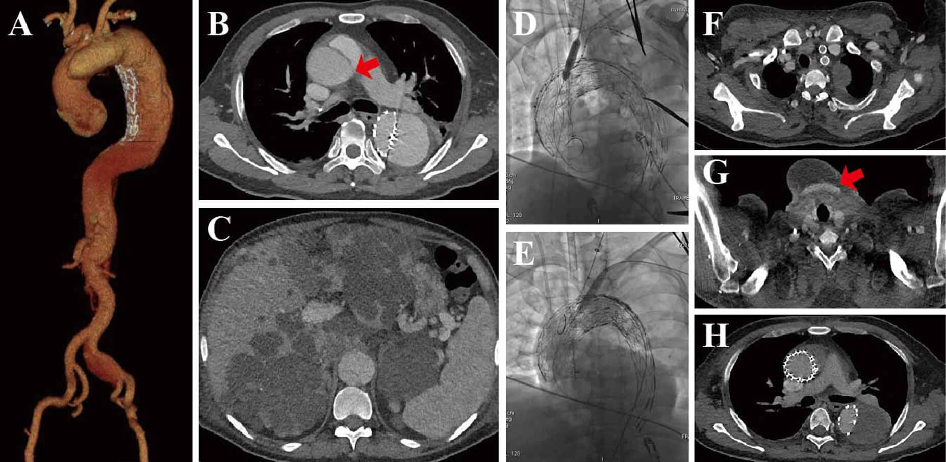
A 47-year-old male with chest and back unbearable pain, shortness of breath and diaphoresis for 2 days was transferred to our hospital for further treatment. He was diagnosed with type B aortic dissection 2 year ago and received thoracic endovascular aortic repair (TEVAR) at the local hospital. He was suffered with polycystic liver, polycystic kidney, chronic anemia, and chronic renal failure. Computed tomography angiography (CTA) scan revealed retrograde type A aortic dissection (Figure 1A), the aortic tear was located at the ascending aorta trunk (Figure 1B and video), and CTA showed he has polycystic liver and polycystic kidney (Figure 1C). Together with the patient’s clinical features, this patient was not suitable for thoracotomy surgery by consultation of Multidisciplinary teamwork (MDT). Upon our study [1], this case might be accessible using in situ laser stent-graft fenestration during TEVAR.
After complete preoperative assessment, the patient successfully underwent minimally invasive treatment with graft replacement at the ascending aorta and in situ laser stent-graft fenestration of the left carotid artery (Figure 1D) and the left subclavian artery (Figure 1E) and revascularization of the right carotid artery by the open bypass of the left carotid artery-to-the right (Figure 1G) under extracorporeal cerebral circulation protection. He was discharged home in good clinical conditions at day 5 without any complications. Post-operative CTA images demonstrated the patency of the left subclavian artery and the left carotid arteries (Figure 1F) and the bypass bridge (Figure 1G) with favorable aortic remodeling (Figure 1H).
As we have known so far, this is the first case report of in situ laser stent-graft fenestration of retrograde type A dissection during TEVAR in poorly pathological conditions, such as polycystic liver, polycystic kidney, chronic anemia, and chronic renal failure.
Funding
This study was financially supported by the National Natural Science Foundation of China (81601621, 81370423, 81570432, 81600205) and the Natural Science Foundation of Shanghai Science and Technology Committee (Grant No. 134119a2100 and 20124Y132).
Conflicts of Interest
None.
REFERENCES
[1] Jinbao
Qin, Zhen Zhao, Ruihua Wang, et al. “In situ Laser Fenestration Is a
Feasible Method for Revascularization of Aortic Arch During Thoracic
Endovascular Aortic Repair.” J Am Heart Assoc, vol. 6, no. 4, pp.
e004542, 2017. View at: Publisher
Site | PubMed