Received: Mon 25, Nov 2024
Accepted: Sat 07, Dec 2024
Abstract
Rhinoplasty performed with the new piezoelectric system, utilizing ultrasonic vibration and various tip, shapes, and sizes, has enhanced precision, safety, and protection of surrounding tissues. This technique applies minimal pressure to the bone, including mobile bones, significantly reducing the risk of unintended fractures. Wide exposure is necessary to ensure optimal bone visualization, enabling delicate rasping and contouring. The dorsal and lateral aesthetic lines are sculpted and refined with ease and safety using the ultrasonic tip, minimizing deformities and instability. However, surgeons must possess adequate training and skill to effectively operate this advanced technological instrument, which is prone to potential breakage or malfunction.
1. Introduction
Rhinoplasty is among the most intricate cosmetic surgeries, requiring meticulous techniques to balance function and aesthetics. Traditional osteotomy methods often involve significant risks, such as uncontrolled fractures, soft tissue damage, and postoperative deformities. In recent years, the piezoelectric device has emerged as a groundbreaking tool in rhinoplasty, offering surgeons greater precision in bone sculpting. This article explores the benefits, challenges, and clinical outcomes of utilizing piezoelectric instruments in rhinoplasty.
2. Case Presentation
Case 1
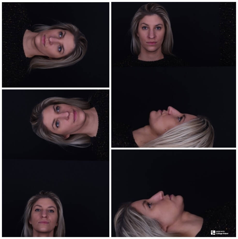
A 34-year-old woman underwent ultrasonic structural open rhinoplasty, presenting with concerns about a broad nose with a dorsal hump, an undefined tip, and a droopy appearance. She described her nose as masculine in appearance. After achieving full exposure of the osseocartilaginous vault, a small bony cap was removed using a piezoelectric rasp tip, carefully preserving the underlying perichondrium. The upper lateral nasal walls were contoured using the same instrument. The cartilaginous hump was reduced by 2 mm while maintaining the integrity of the upper lateral cartilage mucosa. Subsequently, the lateral nasal walls were sculpted using the piezo rasp to narrow the width and decrease the convexity of the nasal bones. Bilateral complete osteotomies, including medial oblique, transverse, and low-to-low cuts, were performed with the piezoelectric device. To refine the tip, a cephalic trim, tip sutures, and a septal extension graft were applied.
Before: Figure 1 illustrates different perspectives of the patient’s nose prior to rhinoplasty.
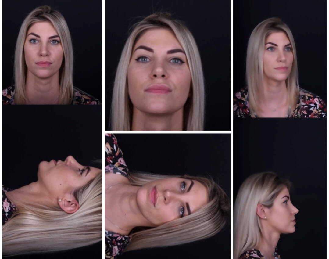
After: Figure 2 displays the outcomes 9 months post-rhinoplasty, showing the refined and balanced nasal contours achieved with the piezoelectric system.
Case 2
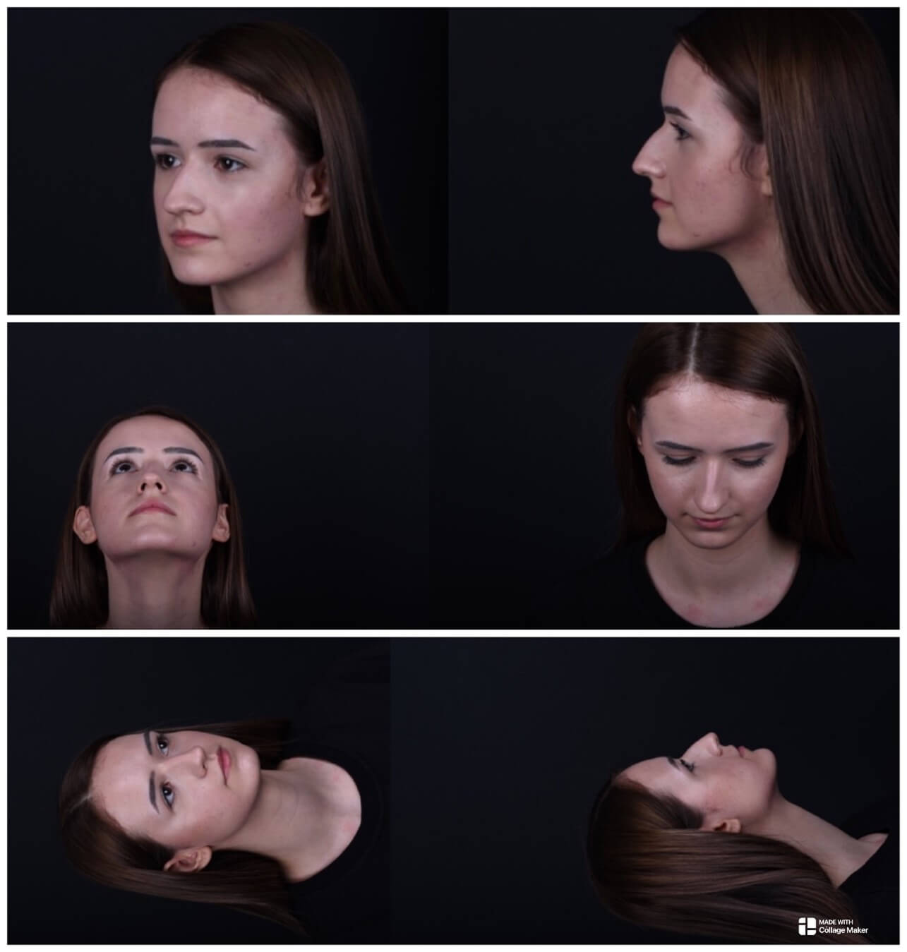
A 23-year-old woman with significant facial asymmetry sought treatment for a crooked, elongated nose with a dorsal hump and droopy tip. She had no history of facial trauma or prior surgery. Additionally, she reported breathing difficulties caused by pronounced S-shaped septal deviation, with cephalic convexity toward the left and caudal convexity toward the right. She underwent a closed low-strip asymmetric let-down preservation rhinoplasty. The procedure included the complete release of the septum from the maxilla and the perpendicular plate of the ethmoid bone. The septal flap was rotated and secured in the midline using a groove custom-crafted in the premaxilla with the piezoelectric device. Complete osteotomies, including transverse, radix, and right low-to-low, along with a left ostectomy, were performed using piezoelectric instruments. The osteocartilaginous vault was shifted en bloc to the left side and medialized. Fine adjustments, including sculpting the sidewalls with the piezoelectric rasp, were also carried out. The tip deformity was addressed with a cephalic trim, tip suturing, and placement of a columellar strut.
Before: Figure 3 depicts various perspectives of the patient’s nose prior to rhinoplasty.
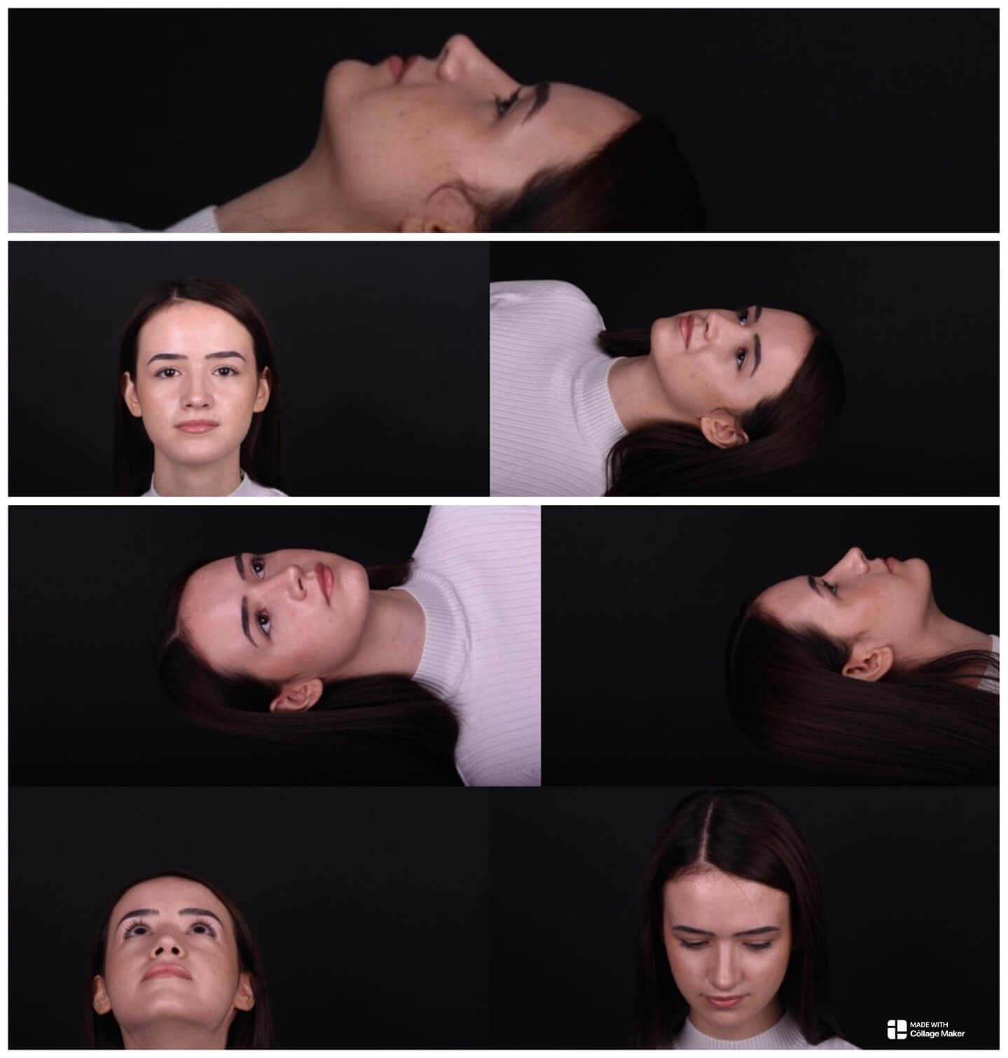
After: Figure 4 highlights the results 10 months post-rhinoplasty, showing the improved symmetry and functional outcomes achieved with the piezoelectric system.
3. Discussion
The introduction of the piezoelectric system into rhinoplasty has significantly enhanced surgical outcomes, particularly in terms of precision, safety, and postoperative healing [1]. This innovative technique leverages high-frequency ultrasonic vibrations transmitted through specialized instrument tips to precisely cut bone while preserving surrounding neurovascular structures and soft tissue [2]. Furthermore, the technique minimizes osteocyte damage, promoting better bone healing during bone harvesting [3]. A substantial reduction in the risk of uncontrolled fractures ensures greater stability during bone mobilization and reshaping [1, 4].
Osteoplasty with the piezoelectric device requires wide and thorough exposure, extending from the keystone area to the radix and involving the release of the pyriform ligament [1]. This exposure allows for precise visualization of deformities and facilitates bony cap removal without creating an open roof or contour irregularities, which are often seen with traditional osteotomy techniques [5]. The ability to use short, angulated saws enables improved rhino-sculpture [6, 7] and safe contouring of the nasal bones to achieve symmetrical narrowing of the dorsum [7].
The piezo device's varied and finely shaped piezoelectric tips enable safe rasping of even mobile bones, applying minimal pressure on targeted areas of the bone's angular surface [8]. With the updated technique, lateral osteotomies are performed first, followed by medial oblique osteotomies if needed [1]. Rasping along the dorsum, particularly on the convex lateral portions of a crooked nose, provides effective narrowing of the dorsum’s aesthetic lines. Refinement of the cephalic border of the medial oblique transition between the bony and cartilaginous vaults helps narrow the nasal pyramid [10, 11]. The piezoelectric device allows for precise lateral osteotomy near the maxillary bone within the nasolabial groove, preventing step deformities that are often palpable and visible. While this approach may be slower than older methods, it is considerably safer [10, 11]. The device also allows for lowering and reshaping the dorsal aesthetic lines without excessive pressure.
To prevent postoperative re-deviation, the piezoelectric drill tip can be used to create a precise hole in the nasal spine for better septal fixation with sutures [7]. Stability for a deviated caudal septum can be further enhanced using the "swinging door" technique, which involves dissecting the cartilage along the maxillary crest and repositioning it over the nasal spine for a straighter alignment [12].
Despite its advantages, the piezoelectric device is not without limitations. Like any advanced technology, it is prone to breakage or malfunction and requires skilled and well-trained surgeons to ensure its effective use. Additionally, the system is more expensive than conventional tools. The wide exposure required for the technique, including skin elevation and ligament release, may result in increased postoperative edema, bruising, and potential external scarring compared to traditional approaches [13].
4. Conclusion
Piezoelectric surgery offers a precise, aesthetic, and safe approach to rhinoplasty, redefining surgical standards with its ability to minimize trauma and improve outcomes. While challenges remain, including cost, training requirements, and device fragility, the advantages in precision and postoperative recovery make it a compelling alternative to traditional osteotomy. By advancing both aesthetic and functional results, this technology provides surgeons with a transformative tool for achieving more natural-looking and predictable outcomes.
Acknowledgements
Figures 1-4 provided by Dr. Witold Waskiewicz.
REFERENCES
[1] Olivier Gerbault, Rollin K Daniel,
Aaron M Kosins “The role of piezoelectric instrumentation in rhinoplasty
surgery.” Aesthet Surg J, vol. 36, no. 1, pp. 21-34, 2016. View
at: Publisher
Site | PubMed
[2] Philippe Hennet “Piezoelectric Bone
Surgery: A Review of the Literature and Potential Applications in Veterinary
Oromaxillofacial Surgery.” Front Vet Sci, vol. 2, pp. 8,
2015. View at: Publisher
Site | PubMed
[3] Lobna Abdel Aziz Aly “Piezoelectric
surgery: Applications in oral & maxillofacial surgery.” Faculty
Dental Journal, vol. 4, no. 2, pp. 105-111, 2018. View
at: Publisher Site
[4] Olivier Gerbault “Ultrasonic
Rhinoplasty and Septoplasty for Dorsum Preservation and for Dorsum Structural
Reconstruction.” Facial Plast Surg Clin North Am, vol. 31, no.
1, pp. 143-154, 2023. View at: Publisher Site | PubMed
[5] Meysem
Yorgun, Erdinç Çekiç, Ali Evlice “Mobile Bony Cap (Meysem Yorgun Technique): An Innovative
Technique in Preservation Rhinoplasty for Crooked Nose Deformity.” Plast
Reconstr Surg Glob Open, vol. 11, no. 4, pp. e4919, 2023. View
at: Publisher Site | PubMed
[6] Pablo Casas-Rodera, Rubén Perez Parra
“Ultrasonic Rhinoplasty: Osteotomies and Dorsum Refinement.” Rhinoplasty
Archive,
2020.
[7] Nevzat
Demirbilek, Cenk Evren, Mustafa Çelik “Piezoelectric Drilling Hole Technique in
Septal Fixation.” J
Craniofac Surg, vol. 30, no. 5, pp. 1544-1548, 2019. View at: Publisher Site | PubMed
[8] Edmund A Pribitkin, Leela S Lavasani,
Carol Shindle, et al. “Sonic rhinoplasty: sculpting the nasal dorsum with the
ultrasonic bone aspirator.” Laryngoscope, vol. 120, no.
8, pp. 1504-1507, 2010. View at: Publisher Site | PubMed
[9] Giancarlo
Tirelli, Margherita Tofanelli, Federica Bullo, et al. “External osteotomy in
rhinoplasty: Piezosurgery vs osteotome.” Am J Otolaryngol, vol. 36, no. 5, pp. 666-671,
2015. View at: Publisher Site | PubMed
[10] Abdülkadir
Göksel, Priyesh N Patel, Sam P Most “Piezoelectric Osteotomies in Dorsal
Preservation Rhinoplasty.” Facial Plast Surg Clin North Am, vol. 29, no. 1, pp. 77-84, 2021. View at: Publisher Site | PubMed
[11] Qian Jiang, Yating Qiu, Chi Yang, et
al. “Piezoelectric Versus Conventional Rotary Techniques for Impacted Third
Molar Extraction: A Meta-analysis of Randomized Controlled Trials.” Medicine
(Baltimore), vol. 94, no. 41, pp. E1684, 2015. View at: Publisher Site | PubMed
[12] N J Pastorek, D G Becker “Treating
the caudal septal deflection.” Arch Facial Plast Surg, vol.
2, no. 3, pp. 217-220, 2000. View at: Publisher Site | PubMed
[13] Ankur Khajuria, Ada M Krzak, Rohin K Reddy, et al. “Piezoelectric Osteotomy versus Conventional Osteotomy in Rhinoplasty: A Systematic Review and Meta-analysis.” Plast Reconstr Surg Glob Open, vol. 10, no. 11, pp. e4673, 2022. View at: Publisher Site | PubMed